
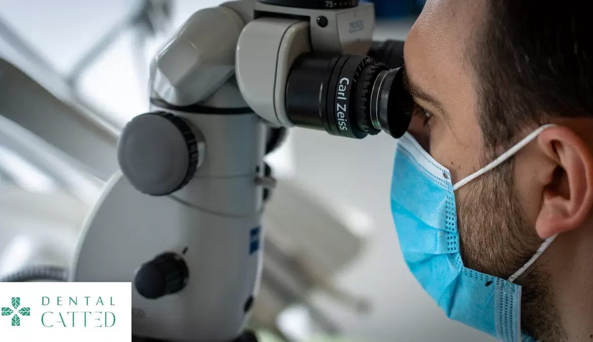
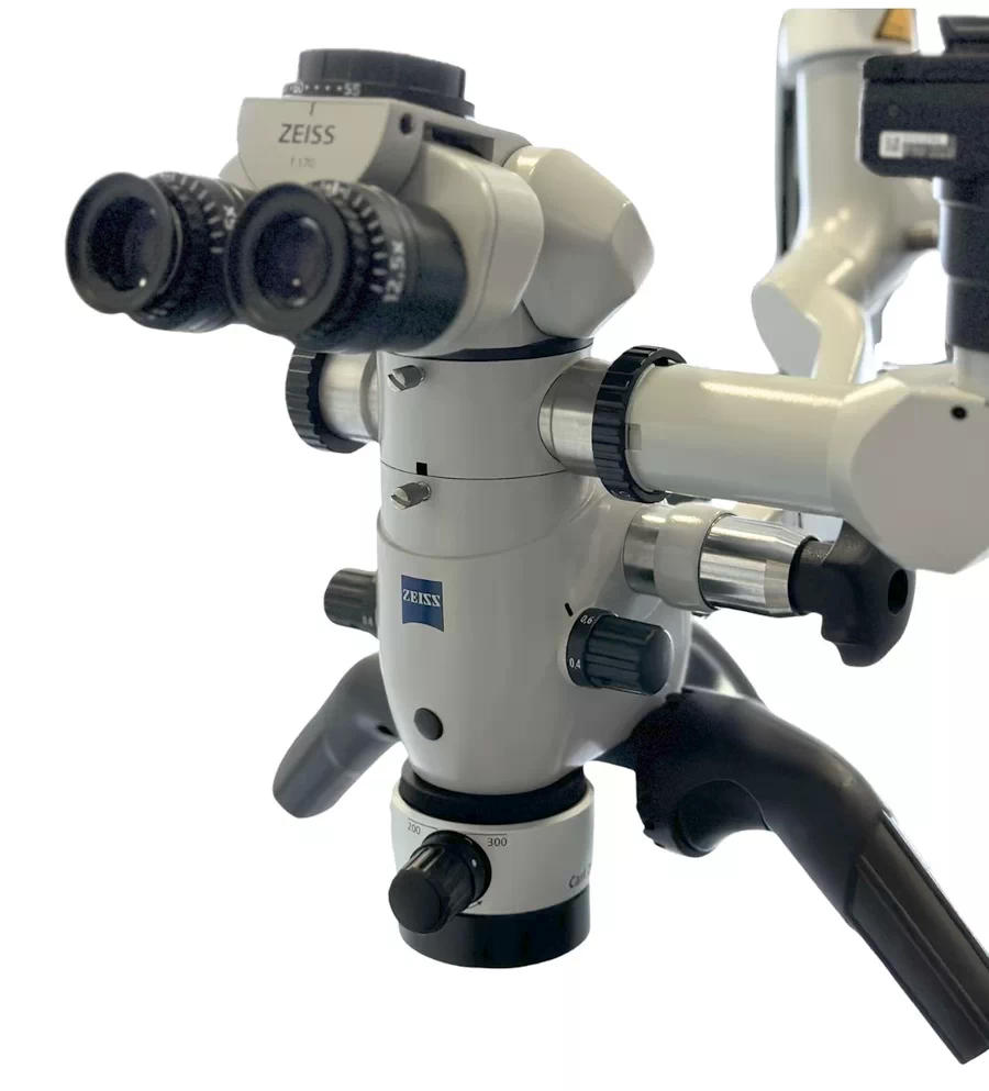
An unsuccessful root canal treatment can cause recurring pain and complications, but modern technology offers precise ways to save the affected teeth. In this detailed guide we look at why root canal treatment fails, the role of the dental microscope and the stages of effective treatment, to help you understand how you can benefit from an advanced approach. At Dental CATTED, we use the ZEISS OPMI Pico microscope to turn endodontic procedures into high-precision treatments, improving the chances of success even in complicated cases.
Common reasons why root canal treatment fails
Root canal treatment involves cleaning out the infection inside the tooth, but failures happen for various reasons:
- Complex root canals missed during the initial procedure.
- Bacterial reinfection due to incomplete sealing.
- Filling materials that break down over time.
- Later trauma or poor oral hygiene.
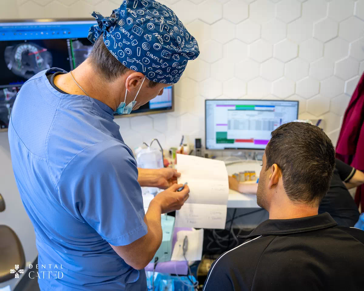
Identifying the causes through advanced diagnosis is the first step toward a solution. For example, root anatomy varies greatly from one patient to another – some teeth have up to four or five hidden, curved or branching canals, which cannot be detected without high magnification. Incomplete sealing lets bacteria back in, leading to chronic inflammation, and traditional materials can shrink or wear away under the effect of saliva and chewing forces. Poor hygiene makes things worse through plaque buildup, turning an initially good treatment into a long-term failure. With 3D X-rays and detailed analysis, we can identify the exact problem and plan a targeted treatment, avoiding unnecessary extractions.
Signs of failure and when to seek help
If you have had root canal treatment and symptoms appear, do not ignore them:
- Persistent pain or throbbing in the area of the tooth.
- Swelling, sensitivity to pressure or to extreme temperatures.
- Recurring abscesses or gum fistulas.
- Discoloration of the tooth or an unpleasant smell.
These signs suggest that a professional reassessment is needed. Throbbing pain, for example, often indicates an active infection pressing on the surrounding nerves, while swelling can signal an abscess spreading toward the soft tissues. Sensitivity to heat or cold persists if remnants of infected tissue have not been completely removed. Gum fistulas can also form – “tunnels” through which pus reaches the surface, causing a characteristic smell. A tooth turning gray or brown is the result of untreated internal necrosis. Ignoring these signals can lead to tooth loss or even systemic infections; that is why a prompt consultation with an endodontist can prevent major complications and help restore your oral health in time.
The advantages of the dental microscope in endodontics
The dental microscope provides magnified visibility, turning procedures into precise treatments:
- It detects hidden or branching canals that are invisible to the naked eye.
- It allows thorough cleaning, reducing the risk of reinfection.
- It makes minimally invasive treatment possible, preserving more healthy tissue.
- It improves the accuracy of sealing for long-lasting results.
This technology is essential for complex cases. With up to 25x magnification and coaxial illumination, the ZEISS OPMI Pico microscope reveals microscopic details such as fine cracks or lateral canals that would otherwise go undetected. Cleaning becomes more thorough, and bacteria and necrotic tissue can be removed more effectively from hard-to-reach areas, which can increase the chances of the treatment succeeding. The minimally invasive approach means smaller openings, less postoperative pain and preservation of the natural tooth structure, which is essential for long-term strength. Precise sealing with biocompatible materials such as thermoplastic gutta-percha creates a tight barrier against reinfection, providing results that can last for years and improving the patient’s quality of life.
The stages of microscope-assisted treatment
The process is structured for comfort and efficiency:
- Initial diagnosis: We use 3D X-rays for a detailed image.
- Local anesthesia: Helps keep discomfort to a minimum.
- Access and cleaning: The microscope guides the precise removal of the infection.
- Sealing: Biocompatible materials for long-term protection.
- Final restoration: Fitting an aesthetic crown to restore function.
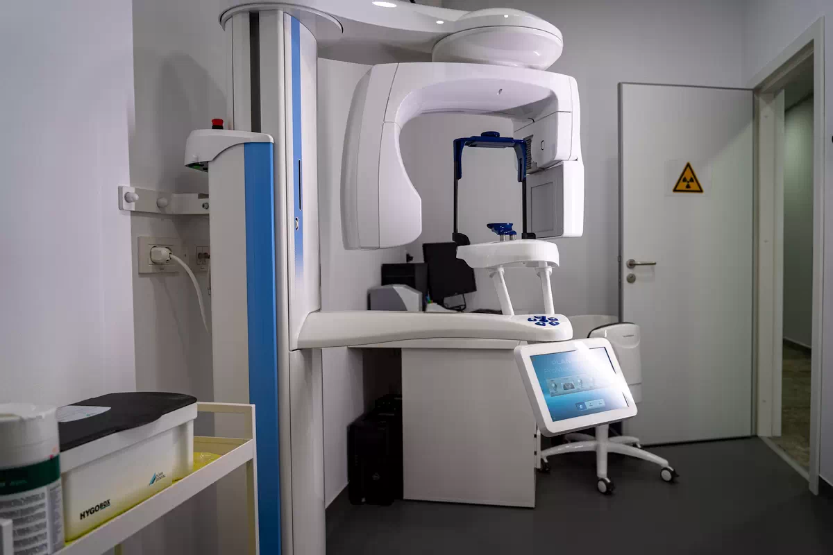
Recovery is usually quick, with clear aftercare instructions. Treatment starts with a CBCT (cone beam computed tomography) scan, which gives a three-dimensional view of the roots and identifies hidden anomalies. Under local anesthesia, the procedure is usually well tolerated, even during longer sessions. Under the microscope, the dentist navigates the canals with fine instruments, removing the infection millimeter by millimeter and irrigating with antibacterial solutions. Sealing uses heat-based techniques for a precise fit, and the final crown – in ceramic or zirconia – restores appearance and chewing strength. After treatment, patients receive personalized advice: avoid hard foods for 24–48 hours, brush gently and attend regular checkups. Most return to their normal activities the same day, with a healthy smile.
Book an appointment now!
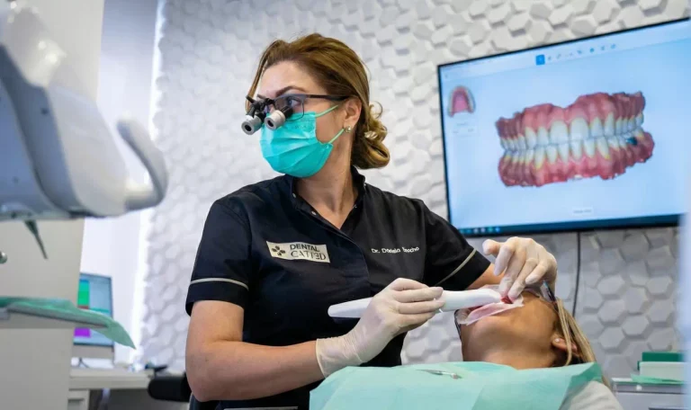
At Dental CATTED, we integrate the ZEISS OPMI Pico microscope into our endodontic services, offering solutions tailored to each patient. Our team uses equipment such as the ZEISS OPMI Pico microscope and the Planmeca ProMax, which help make treatment precise and as comfortable as possible.
If you suspect a failed treatment, contact us at +40 733 22 88 28 or fill out the contact form for a detailed assessment.
Many teeth with a failed root canal treatment can be kept through careful retreatment. Book a consultation to find out what options you have in your case.